
Researchers from the team led by Claudia Sissi of the Department of Pharmaceutical Sciences at the University of Padova have contributed to the study of the structure of DNA, commonly known as a double helix. The goal of this study was to explain why there are two molecular “knots” on two strands of a short segment of DNA.

My note: I want to make it clear that this article is not intended to be a standalone scientific study. Through this blog, I hope to carefully share scientifically relevant information. If you’d like to explore this topic in greater depth, I invite you to refer to the links to the scientific research that I’m sharing here so you can analyze all the results.
For years, the scientific community maintained the opposite—that such a thing was impossible.
The structure of DNA is quite dynamic: it can unfold, fold, and take on different shapes; as a result, we can speak of “knots” that influence the way genetic information is read and used by the cell. Two specific, unconventional structures, known as G-Quadruplex and i-Motif, can coexist within the same DNA segment. Guanine-rich sequences can fold into G-quadruplexs (G4s), while the complementary strand adopts potentially i-Motif (iM) arrangements.

Photo from Unraveling G‐Quadruplex and i‐Motif Coexistence Within a Double‐Stranded DNA – Auricchio – Angewandte Chemie International Edition – Wiley Online Library. The relative thermodynamic profiles of G-quadruplex (G4), i-Motif (iM), and the duplex counterpart tune the simultaneous G4 and iM formation within a double-stranded (ds) DNA.
G4s is a tetra-helical arrangements driven by the pairing of four guanines through Hoogsteen hydrogen bonds. At most G4-forming sites, the complementary C-rich strand can potentially fold into i-Motif (iM), antiparallel tetra-helical structures held together by intercalated hemi-protonated C:C base pairs. Since cytosine pK is ≈4.6, originally iMs were not considered as physiologically relevant. However, in vitro, by increasing the number of C:C base pairs and/or by applying molecular crowding conditions to simulate the nuclear environment, it was possible to observe iM formation by C-rich genomic sequences also at pH close to the physiological one.
Researchers have designed an experimental DNA model that allows them to observe the behavior of these two unconventional structures under controlled and reversible conditions—conditions very similar to those found in cells, using an integrated suite of spectroscopic, electrophoretic, calorimetric, and scattering techniques, including UV absorption spectroscopy (UV), circular dichroism (CD), polyacrylamide gel electrophoresis (PAGE), differential scanning calorimetry (DSC), and small-angle x-ray scattering (SAXS). Subsequently, researchers modified the central segment to meet specific requirements that emerged during the project development. They involved both nucleotide length (27 or 33 nt) and sequence composition (27, 27*, 33*, and 33 m), applied to the G-rich, the C-rich, or both strands. Overall, this allowed us to mimic both a canonical and an unwound B-DNA, where the reduced helical stability is expected to boost the folding of noncanonical structures. All full-length constructs and isolated domains have been comprehensively characterized. By merging the acquired results, we addressed at the molecular level the G4/iM co-localization, providing the thermodynamic parameters that support their simultaneous formation.
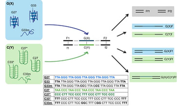
Photo from Unraveling G‐Quadruplex and i‐Motif Coexistence Within a Double‐Stranded DNA – Auricchio – Angewandte Chemie International Edition – Wiley Online Library. Schematic representation of the modular structural construct used in this study. The sequences of the selected G- and C-rich domains that differ in length (27 or 33 nts) and composition (27*, 33*, and 33 m)—overall summarized as G(X) and C(Y)—are reported in the central panel. Residues in bold indicate sequence modifications. Ff1 and Ff2 are pairs of fully complementary sequences added at the terminals of the G(X) and C(Y) strands.
From these studies, the occurrence of iM and G4 at overlapping sites emerged, thus suggesting that both these structural modules can work as regulatory elements of related biological processes, either individually or through fine-tuning of protein recruitment. n addition, early biochemical and single-molecule studies reported simultaneous G4- and iM-folding only when the two tetra-helices were located out-of-phase at a proper distance along the complementary strands, pointing to their mutual exclusion due to steric hindrance. However, it is worth noting that the present work focused only on a single GC-rich pair. Although the selected G-rich telomeric sequence can fold into diverse G4 topologies, the behavior of G4 and iM modules with different constrained topologies, as well as the impact of their possible structural rearrangements when folding within a longer DNA fragment, needs to be considered.
The study has important implications for human health. Certain diseases are associated with the abnormal presence of genetic sequences capable of forming these structures. The presence of too many or too few of these nodes can disrupt the proper functioning of the cell, altering the way genetic information is interpreted.
You can find what I’m talking about at the following link: Unraveling G‐Quadruplex and i‐Motif Coexistence Within a Double‐Stranded DNA – Auricchio – Angewandte Chemie International Edition – Wiley Online Library.
Thank you for reading